Introduction
Essential thrombocythemia (ET) is a clonal myeloproliferative neoplasm (MPN) characterized by sustained thrombocytosis and megakaryocytic proliferation driven by acquired mutations affecting thrombopoietin receptor signaling.1 According to the 2022 International Consensus Classification (ICC), ET belongs to the group of BCR::ABL1-negative MPNs alongside polycythemia vera (PV), primary myelofibrosis (PMF) and MPN-unclassifiable (MPN-U). Hereditary forms are rare.1,2
The molecular landscape of ET has been shaped by the discovery of three principal driver mutations. The identification of JAK2 V617F in 2005 established constitutive JAK–STAT signaling as a central pathogenic mechanism, followed by the discovery of MPL mutations in 2006 and CALR mutations in 2013. Together, these alterations account for approximately 85–90% of cases, while the remaining 10–15% of patients have so-called triple-negative disease.3
ET is among the most common chronic MPNs, with an annual incidence of 0.38–1.7 cases per 100,000 person-years, a female predominance and a median age at diagnosis of 58–59 years.1
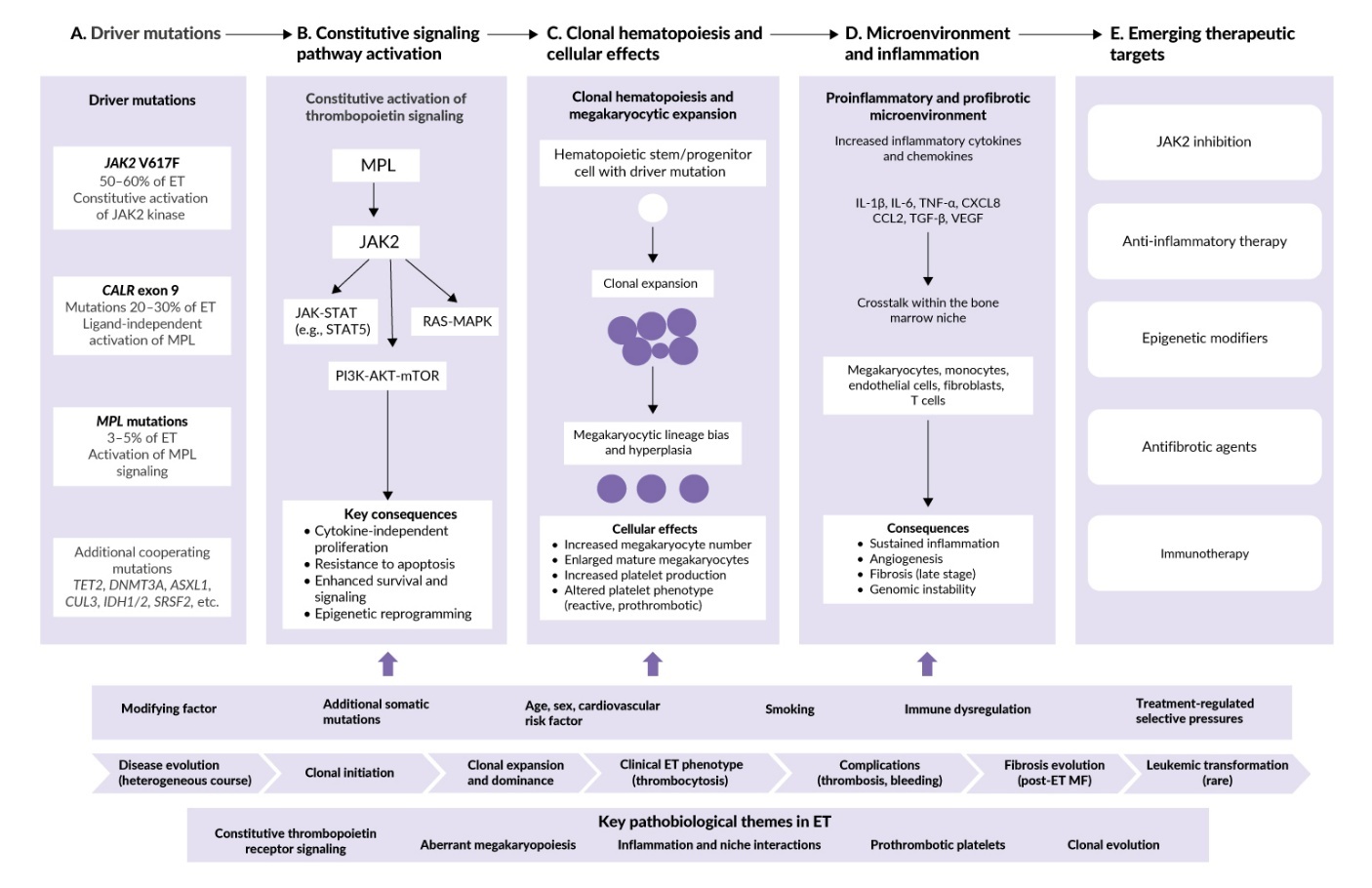
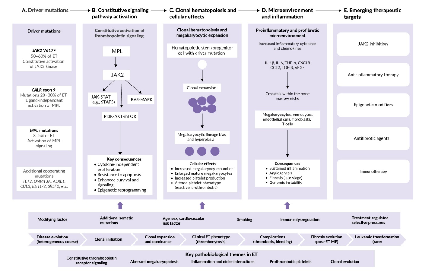
Advances in molecular characterization have refined diagnosis and prognosis in ET and driven the development of targeted therapies, opening new treatment horizons. This review outlines the current understanding of ET pathogenesis, diagnostic approaches, risk stratification and evolving therapeutic strategies, with an integrated overview of pathogenesis and emerging therapeutic targets presented in Figure 1.
Mechanistic and inflammatory models in the pathogenesis of essential thrombocythemia
Molecular drivers
Recent molecular and translational studies have revealed that essential thrombocythemia is not a single homogeneous disorder but rather a spectrum of molecularly defined disease entities, in which distinct driver mutations and associated co-mutation patterns determine disease biology, clinical presentation, thrombotic risk, therapeutic responsiveness and the risk of myelofibrotic transformation.4
All three canonical driver mutations – JAK2 V617F, CALR and MPL – converge on constitutive activation of the JAK-STAT, PI3K and RAS-MAPK signaling pathways, bypassing the normal requirement for thrombopoietin.5
JAK2 V617F and CALR mutations drive MPN pathogenesis through distinct mechanisms. Heterozygous JAK2 V617F forms heterodimers with wild-type JAK2, preferentially activating STAT1 over STAT5 and inducing aryl hydrocarbon receptor (AhR) to bias megakaryocyte-erythroid progenitor differentiation toward megakaryopoiesis.6
In contrast, CALR mutations generate a novel positively charged C-terminal neoantigen that lacks the endoplasmic reticulum retention signal. This mutant CALR is both secreted extracellularly and trafficked to the cell surface in complex with immature MPL, making the neoantigen accessible for immunotherapeutic targeting.7 Both type 1 (52-bp deletion) and type 2 (5-bp insertion) CALR mutations are gain-of-function alterations that activate MPL and trigger distinct cellular stress response programs. These differences account for variation in clinical phenotype and prognosis, with type 1 mutations being more pathogenic and associated with greater thrombocytosis, earlier stem cell expansion and increased myelofibrotic transformation.8
MPL (myeloproliferative leukemia virus oncogene) encodes the thrombopoietin receptor and serves as a central regulator of megakaryopoiesis and hematopoietic stem cell self-renewal.4 Discovered one year after JAK2 V617F, MPL mutations remain the least studied class of MPN driver mutations, likely due to their low incidence (3–5% of cases). However, evidence suggests that the thrombotic risk is comparable to or potentially exceeds that of JAK2-mutated disease. The W515 residue in the transmembrane-juxtamembrane domain is essential for ligand-independent oncogenic activation and represents a potentially actionable therapeutic target through selective disruption of pathologic MPL dimerization.9
Triple-negative essential thrombocythemia (TN-ET), accounting for approximately 10% of World Health Organization (WHO)-defined ET, represents a misleading designation: beneath the absence of canonical JAK2, CALR and MPL drivers lies substantial molecular complexity. Cryptic JAK2 V617F clones with very low variant allele frequency (VAF <1%) are detectable only by ultrasensitive techniques, including droplet digital PCR and ultra-deep next-generation sequencing.10 The molecular landscape of TN-ET is further defined by non-canonical MPL variants, clonal hematopoiesis of indeterminate potential (CHIP)-associated mutations (TET2, DNMT3A, ASXL1) and additional lesions in epigenetic regulators and signaling genes (EZH2, IDH1/2, NFE2, SH2B3).11
Triple-negative ET is generally associated with a lower thrombotic risk and a lower risk of fibrotic transformation.12 However, co-mutations modify these outcomes: TET2 mutations may increase thrombotic risk, while ASXL1 mutations confer inferior survival and increased thrombotic risk across MPN subgroups. This molecular heterogeneity translates into clinical unpredictability, variable prognosis and differential treatment responses, particularly to interferon therapy.
While these driver mutations provide the molecular ignition for ET pathogenesis with distinct clinical phenotypes by molecular subgroup (Table 1), clonal expansion and disease evolution depend critically on a second component – chronic inflammation.
Inflammatory microenvironment
Established concepts
Regardless of the driver mutation, ET demonstrates activation of NF-κB, MAPK and JAK-STAT signaling pathways that drive aberrant megakaryopoiesis and create a proinflammatory bone marrow microenvironment.13 Toll-like receptor (TLR) signaling, particularly through TLR2, is elevated in ET patients and correlates with JAK2 V617F mutation status and leukocytosis. TLR activation drives production of IL-1β, IL-6, TNF-α and interferons in the bone marrow.14 Monocytes from MPN patients exhibit defective IL-10-mediated negative feedback, resulting in unrestrained TNF-α production after TLR activation.14 This inflammatory microenvironment selectively favors expansion of JAK2 V617F-positive cells over normal hematopoiesis.15
Beyond promoting clonal expansion, inflammatory signaling directly shapes disease phenotype through effects on megakaryopoiesis and platelet function. Supporting this concept, Yañuk et al. demonstrated that plasma derived from ET and myelofibrosis patients differentially regulates megakaryopoiesis through distinct cytokine profiles.13 In parallel, Oyarzún et al. showed that TLR-mediated platelet hyperreactivity in ET is associated with enhanced P-selectin translocation, increased platelet-neutrophil aggregate formation and altered α-granule release, establishing a direct mechanistic link between innate immune activation and thromboinflammation in ET.16
Emerging and speculative findings
TLR signaling triggers production of inflammatory cytokines including IL-6 and IL-8, which further amplify inflammation through additional immune pathways.17 However, these mechanisms have primarily been characterized in myelofibrosis models, and ET-specific validation remains limited.
The NLRP3 inflammasome has emerged as a potential therapeutic target based on preclinical MPN studies. In JAK2 V617F-mutant mouse models, NLRP3 deletion prevents IL-1β and IL-18 release and reduces thrombocytosis, splenomegaly and bone marrow fibrosis. Pharmacologic NLRP3 blockade reverses established disease features in these models.18 However, these findings are derived entirely from myelofibrosis-focused preclinical studies. NLRP3 inflammasome activation and its therapeutic relevance specifically in ET remain unvalidated.
Although inflammatory signaling is increasingly recognized as a contributor to ET pathogenesis, therapeutic strategies directly targeting these pathways remain unexplored in clinical trials.
Clinical manifestations and diagnostic evaluation
Diagnosis requires integration of clinical, molecular and bone marrow findings. Reactive thrombocytosis must be excluded first, as secondary causes including infection, inflammation, iron deficiency, postoperative states or splenectomy account for most cases of thrombocytosis. Detection of JAK2, CALR or MPL mutations supports the diagnosis, whereas mutation-negative cases require exclusion of BCR::ABL1-positive chronic myeloid leukemia and, when appropriate, expanded myeloid molecular testing.19
Bone marrow biopsy remains essential for distinguishing ET from prefibrotic primary myelofibrosis (pre-PMF), overt myelofibrosis, myelodysplastic/myeloproliferative neoplasms and masked polycythemia vera. This distinction carries important prognostic implications: pre-PMF is associated with inferior overall survival and higher rates of fibrotic and leukemic progression compared with ET.20
Key diagnostic differences among thrombocytosis-presenting MPNs are summarized in Table 2.
The clinical burden of essential thrombocythemia extends beyond thrombocytosis to include fatigue, vasomotor symptoms, erythromelalgia, pruritus and splenomegaly, all of which may impair the quality of life. Major complications include arterial and venous thrombosis, hemorrhage, pregnancy-related morbidity and progression to myelofibrosis or acute leukemia.1 The main clinical manifestations and reported frequencies are summarized in Table 3.
From thrombosis to survival: Modern risk stratification in essential thrombocythemia
Risk stratification in ET has evolved from a thrombosis-focused assessment toward an integrated clinical and molecular prognostication. The revised International Prognostic Score for Essential Thrombocythemia (IPSET)-Thrombosis score remains the standard tool for thrombotic risk prediction, whereas survival models, subsequently refined by Tefferi et al., incorporate clinical and genomic variables to improve long-term outcome prediction. Molecular profiling has further enhanced prognostication by identifying adverse mutations associated with inferior survival and increased risk of disease progression.24 Key prognostic tools are summarized in Table 4.
Although IPSET-Thrombosis and IPSET-Survival remain practical and widely validated tools for routine clinical care, they rely predominantly on clinical variables and driver mutation status. Newer prognostic models expand on these frameworks by incorporating high-risk somatic mutations (e.g., ASXL1, SRSF2, U2AF1, TP53), measures of clonal complexity and, increasingly, inflammatory biomarkers. These additions may improve patient selection and risk stratification, thereby enabling more individualized monitoring, optimizing the selection of candidates for cytoreductive or investigational therapies and facilitating the implementation of biomarker-guided treatment algorithms.
Current treatment options
Current treatment strategies in ET remain largely risk-adapted and are primarily directed toward thrombosis prevention, hematologic control and symptom management. Hydroxyurea remains the preferred first-line cytoreductive therapy for most high-risk patients, whereas anagrelide and ruxolitinib are generally reserved for selected patients with intolerance, resistance or persistent symptom burden.25–28 Aspirin continues to represent the cornerstone of antithrombotic management in appropriately selected patients, although caution is warranted in the setting of extreme thrombocytosis and acquired von Willebrand syndrome. Despite substantial advances in the understanding of ET biology, currently available non-interferon therapies have limited effects on clonal evolution and disease progression.28 Increasing insights into the molecular and inflammatory mechanisms underlying ET have therefore stimulated the development of biologically targeted and potentially disease-modifying approaches. Key studies supporting established non-interferon therapies are summarized in Table 5, while interferon-based and emerging therapeutic strategies are discussed separately.
Interferon-based therapy and disease modification
Through their ability to target malignant hematopoietic stem cells, reduce mutant allele burden and modulate antitumor immunity, interferons achieve hematologic response rates of approximately 80–86% with sustained molecular responses in both JAK2- and CALR-mutated disease. Randomized studies MPD-RC 112 and SURPASS-ET further established interferon-based therapy as an effective treatment option, particularly in patients with hydroxyurea-resistant or -intolerant ET.29,30 Notably, peginterferon alfa-2a was approved in Europe based on the MPD-RC 112 study, and ropeginterferon alfa-2b is approved globally for polycythemia vera and in some regions for ET; however, no interferon formulation is currently approved in Switzerland for ET treatment.
Newer formulations, particularly ropeginterferon alfa-2b, have demonstrated improved tolerability compared with earlier pegylated interferons. Key studies evaluating interferon efficacy are summarized in Table 6.
Importantly, molecular responses appear to be influenced by the co-mutation status. The presence and timing of specific mutations warrant distinct therapeutic considerations. Pre-existing DNMT3A mutations at treatment initiation should not preclude interferon use, whereas treatment-emergent DNMT3A clones during therapy are associated with hematologic resistance and treatment failure, supporting serial molecular monitoring to identify patients requiring treatment modification.32 While TET2 mutations may reduce molecular response to interferon in some studies, data remain limited and conflicting, and current evidence does not support withholding interferon based on TET2 status alone. ASXL1-mutated patients warrant careful consideration given their adverse prognosis, although ASXL1 is not included in the Mutation-Enhanced International Prognostic System for Essential Thrombocythemia (MIPSS-ET) prognostic model, which instead incorporates SRSF2, SF3B1, U2AF1 and TP53.33 Despite these emerging molecular insights, optimal molecular monitoring strategies and the clinical significance of treatment-emergent clonal evolution remain unresolved.
Emerging therapies and investigational approaches
Increasing evidence suggests that MPNs originate from long-lived hematopoietic stem cell (HSC) clones, making eradication of the malignant stem cell compartment a major therapeutic objective. Interferon remains the only therapy associated with molecular responses, while investigational strategies targeting epigenetic regulation, telomere maintenance and mutation-specific mechanisms are under evaluation as potential clone-directed approaches.
Bomedemstat, an LSD1 inhibitor targeting HSC self-renewal and megakaryocytic differentiation through epigenetic modulation, demonstrated disease-modifying activity across JAK2-, CALR- and MPL-mutated ET with durable platelet control and reductions in driver mutation burden.34
The telomerase inhibitor imetelstat similarly produced hematologic and molecular responses in refractory ET, although subsequent development has focused on myelodysplastic syndromes and myelofibrosis.35
Advances in understanding mutation-specific biology have enabled the development of targeted therapies designed to directly suppress malignant clones. The monoclonal antibody INCA033989 represents the first mutation-selective therapy directed against the mutant CALR–MPL complex. Preclinical studies demonstrated selective elimination of CALR-mutated hematopoietic cells while sparing normal hematopoiesis, and interim clinical data indicate encouraging hematologic and molecular responses with reductions in platelet counts and mutant allele burden.36 Not all precision approaches translate across MPN subtypes: antifibrotic approaches such as AVID200, a selective TGF-β1/3 trap, show activity in myelofibrosis but have limited applicability to ET.37
The immunogenic nature of mutant CALR has opened up the possibility of neoantigen-directed immunotherapy. Early peptide vaccine studies by Grauslund et al. and Holmström et al. targeting the mutant CALR C-terminus demonstrated the induction of CALR-reactive T-cell responses but limited clinical efficacy, likely reflecting immune suppression within the bone marrow microenvironment.44,45 Subsequent strategies combining neoantigen vaccination with checkpoint modulation, including VAC85135 plus ipilimumab, are currently undergoing clinical evaluation.
Adoptive cellular therapies represent the most advanced extension of this concept. Recently developed mutant CALR-directed chimeric antigen receptor (CAR) T cells selectively eliminated CALR-mutated clones in preclinical xenograft and organoid models while preserving normal hematopoiesis, providing proof of concept for potentially curative stem cell–directed immunotherapy in CALR-mutated ET, although this approach has not yet entered clinical testing.46
Conclusions
Essential thrombocythemia is increasingly recognized as a biologically heterogeneous immunoinflammatory stem-cell neoplasm in which mutation-specific clonal architecture, chronic inflammation, immune dysregulation, and microenvironmental interactions collectively shape disease phenotype and long-term evolution. This understanding has catalyzed a fundamental shift from thrombosis prevention and cytoreduction toward precision therapies targeting clonal hematopoiesis, restoring immune surveillance and exploiting mutation-specific vulnerabilities.
Critical questions remain: Should treatment target the malignant clone, the inflammatory microenvironment or both simultaneously? How should patients be selected for emerging precision interventions? As increasingly sophisticated therapies advance from preclinical models to clinical trials, ET management will require the integration of molecular profiling, inflammatory biomarkers, immune signatures and serial clonal monitoring to enable individualized therapeutic selection. Ultimately, ET should no longer be viewed solely as a disorder of thrombocytosis but rather as a complex stem cell neoplasm requiring integrated strategies to achieve durable disease control.
Conflict of interest
The authors declared that the study was conducted in the absence of any commercial or financial relationships that could be construed as a potential conflict of interest.
Funding
The authors have declared that no financial support was received from any organization for the submitted work.
Author contributions
The authors have created and approved the final manuscript.
Acknowledgements
The authors thank Mihailo Ratknic, Senior Research Associate at the Institute of Forestry in Belgrade, Serbia, for his valuable assistance with the figure and table preparation.